Look a little closer
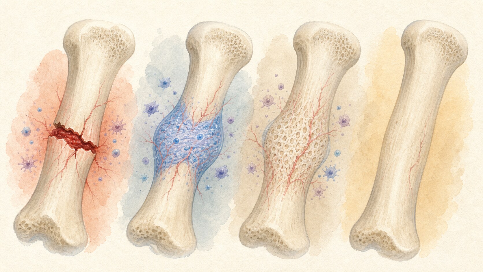
A broken bone does not heal by gluing two fragments together. Bleeding and inflammation establish a repair environment, a cartilage-rich bridge stabilizes the gap, and that bridge is replaced by immature bone. Long-term remodeling then reshapes the repair along the stresses the bone must carry.
Immediately after a fracture, torn vessels bleed into the site and form a clot called a fracture hematoma. Immune cells clear damaged material while releasing signals that recruit progenitor cells and new blood vessels. At this stage, inflammation is not merely an obstacle; it is part of the controlled sequence that starts repair.
Fibrous tissue and cartilage next form a soft callus between the fragments. It is not strong enough for normal loading, but it reduces motion and provides a scaffold for later tissue. Adequate blood supply and a suitable amount of stability are essential if this temporary bridge is to mature.
As the cartilage matrix mineralizes and vessels enter, osteoblasts lay down rapidly produced immature woven bone. The soft callus becomes a hard callus, which may appear as thickening around the fracture on an X-ray. Osteoclasts later remove unnecessary material while osteoblasts build more organized lamellar bone, restoring contour and internal structure.
The mechanical environment determines which healing pattern dominates. Rigid compression with almost no motion can permit primary healing, in which microscopic bone-remodeling units cross the fracture without a large external callus. Controlled relative motion favors secondary healing through cartilage and callus. Casts, plates and other fixation devices do not manufacture bone; they provide the alignment and stability biology needs.
Location, fracture pattern, blood supply and infection influence recovery, as do age, smoking, nutrition and conditions such as diabetes. This does not mean one supplement or stimulation device accelerates every fracture. Less pain, apparent union on an image and recovery of the bone's original strength are also different milestones.
Fractures therefore require diagnosis and follow-up from a qualified clinician. Removing support or increasing weight-bearing without permission can disturb alignment and delay union, while safe movement at the stage recommended by the care team helps joints and muscles recover. Bone healing succeeds through the combination of cellular regeneration and an appropriately protected mechanical environment.
EDITORIAL RESPONSIBILITY
FactosBrain Editorial Desk
The FactosBrain Editorial Desk researched and reviewed this article under our editorial policy. We assess error reports under our corrections policy.